Comprehensive Cardiac Care

To prepare patients prior to being seen at the Orillia Cardiac Care clinic, please review below what you can expect when you’re here to see Dr. Nolan for assessment and when you’re here for Diagnostic Testing.

A medical monitor displaying various vital signs including heart rate and pulse. The screen shows numbers like 139 and 78, with graphical representations of the data below on a printout. The device has several buttons for interaction at the bottom.
Two stethoscopes with shiny metallic diaphragms are arranged on a reflective black surface, with a small red heart placed in the center between them.
Cardiology Follow Up

Your follow-up Cardiology Consultation with Dr. Nolan will allow him to review your progress, assess the results of any recent testing, and make adjustments to your treatment plans as needed.

Cardiology Consultation

Your first visit with Dr. Nolan, your initial Cardiology consultation, is an important step in understanding your heart health. This consultation allows him to assess your cardiac concerns, review your medical history, and begin planning any necessary investigations or treatments.

During your appointment, Dr. Nolan will:

1. Review the referral and any concerns noted your family doctor, ER doctor, and/or specialist

2. Take a detailed history, including your personal and family medical background

3. Discuss any current symptoms you may be experiencing (such as chest pain, shortness of breath, palpitations, dizziness and/or fatigue)

4. Review any prior testing (such as ECGs, blood work results, diagnostic imaging, stress tests and/or any other cardiac related investigations you may have completed)

5. Perform a focused physical examination (support staff will take an updated height, weight, and blood pressure measurements when you are in the office)

6. Discuss the need for further testing, such as echocardiograms, holter monitors, stress tests and other testing if indicated

7. Provide guidance and, if appropriate, begin a management plan for your cardiovascular health

Please bring a list of all current medications (this can easily be obtained from your pharmacist), including over-the-counter supplements, and be prepared to discuss your health history. If you’ve had any recent cardiac testing done outside of our office, please ensure your referring physician has forwarded the results.

This initial visit is an opportunity to begin working together toward a clearer understanding of your heart health and to develop a personalized care plan that meets your needs.

During your follow-up visit, we may:

1. Discuss the results of any recent tests/investigations (such as echocardiograms, Holter monitors, stress tests, lab results, etc)

2. Review your response to medications and/or lifestyle changes

3. Monitor any diagnosed heart conditions

4. Address any new or ongoing symptoms related to your heart

5. Update your care plan based on how your condition has progressed

6. Coordinate any additional testing or referrals, where needed

These visits are a key part of ensuring your heart health is being managed safely and effectively. They give us the opportunity to fine tune your treatment and ensure that any concerns are addressed in a timely manner.

Please bring an updated list of medications to each visit and be prepared to discuss any changes in your symptoms, lifestyle, or overall health

3. Treadmill Stress Test
  • A treadmill stress test is a non-invasive test to assess your exercise capacity, whether your heart is receiving enough blood flow during exercise, and if you abnormal heart rhythms that occur during exercise.

  • There is no special preparation required for this test. Avoid applying cream or lotion to your chest. Take all medications unless advised by physician to stop as some medications can interfere with the test. Bring comfortable clothes to exercise in, running shoes, and a water bottle.

  • The test takes approximately 30-45 minutes. Most patients exercise for between 5 to 15 minutes. You will be on a treadmill and exercise to “stress” the heart. During this you will be hooked up to an ECG machine with stickers on your chest to monitor your heart’s electrical activity during exercise, and your blood pressure will be taken periodically while exercising. The speed will start at a slow walk on a flat surface and every 3 minutes the speed and incline will increase. The test will be stopped when you reach your target heart rate, or sooner if you tell the technologist that you wish to stop.

  • You will have your blood pressure, heart rate and ECG monitored for 5 minutes after you complete the test. You can resume your normal daily activities, including driving, after a treadmill stress test. The results will be reviewed, and a report sent to your doctor.
1. ECG
  • An electrocardiogram (ECG or EKG) is a short recording of the electrical activity of the heart using stickers (called leads) that are placed on the chest. It allows us to detect abnormal heart rhythms associated with heart disease.

  • There is no special preparation required for this test. Wear a loose-fitting top that can easily be removed to facilitate the test. Avoid applying cream or lotion to your chest. You can take all your medications as scheduled.

  • The test takes as little as 15-20 minutes. During the procedure 12 stickers are attached to your chest, arms and legs. Men may need to have their chest hair shaved to allow for better contact. You lie flat and the computer takes a picture of the electrical activity of your heart.

  • You can resume your normal daily activities, including driving, after an ECG. The results will be reviewed, and a report sent to your doctor.

2. Echocardiogram
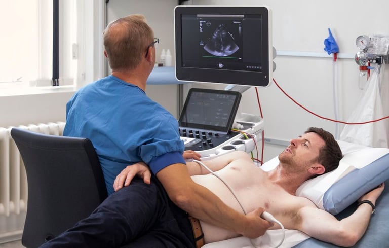
  • An echocardiogram (echo or TTE) is an ultrasound of the heart. It is safe and non-invasive test. Sound waves are used to assess the structure of the heart, as well as the function of the muscle of the heart, valve function and size of the great vessels.

  • There is no special preparation required for this test. Avoid applying cream or lotion to your chest. You will change into a gown prior to the test. You can take all your medications as scheduled.

  • The test takes approximately 45 to 60 minutes. You will lay on a table, on your left side. The sonographer will place a probe on your chest and take images.

  • In some cases, to help improve image quality, we may recommend an injection of an ultrasound enhancing agent. The solution contains microbubbles that improve the image quality and allow better assessment of the heart function. This will require IV insertion.

  • You can resume your normal daily activities, including driving, after an echocardiogram. The results will be reviewed, and a report sent to your doctor.

5. Holter Monitor
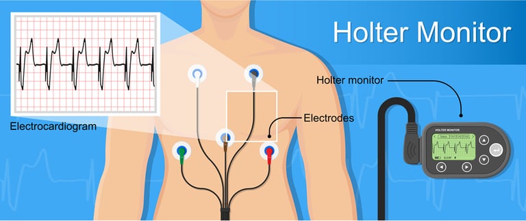
  • A Holter monitor is a wearable device that allows for continuous recording of the electrical activity of the heart for an extended period (24 hours to 7 days).

  • There is no special preparation required for this test. Avoid applying cream or lotion to your chest.

  • The application of the Holter monitor will take approximately 30 minutes. You are fitted with a device with ECG stickers placed on your chest and shown how to wear the device. You then wear the device continuously until you return to the office at the end of the test period. The Holter monitor will monitor your electrical activity while you do your daily activities, and at night. Do not restrict your activities in any way, continue to take your medications as previously scheduled, do not remove the device, and do not get it wet. You will need to keep a diary in which you can record any symptoms experienced including the time and activity level associated with the symptom.

  • You can resume your normal daily activities, including driving, during and after a Holter monitor. The results will be reviewed, and a report sent to your doctor.

4. Stress Echocardiogram
  • A stress echocardiogram (stress echo) is a combination of a treadmill stress test, and an echocardiogram. It is used to assess the function of the heart (or in some cases valves) at rest versus with exercise. It is most often used to look for coronary artery disease (CAD).

  • In preparation, do not eat 3 hours prior to the test. Avoid caffeine (coffee, tea, soft drinks, chocolate) for 6 hours before the test. You may be asked to hold certain medications that slow your heart rate down (beta-blockers, calcium channel blockers) to increase the chances of a successful test. Please confirm with your doctor if you need to hold any of your medications prior to the test. Avoid applying cream or lotion to your chest. Bring comfortable clothes to exercise in, running shoes, and a water bottle.

  • The test takes approximately 30 to 60 minutes. It consists of three parts. First baseline echo pictures of the heart at rest are taken. Then you will get on a treadmill and exercise to “stress” the heart. During this part you will be hooked up to an ECG machine with stickers on your chest to monitor your heart’s electrical activity during exercise, and your blood pressure will be taken periodically while exercising. Once optimal exercise levels have been reached, you will get off the treadmill and quickly move over the to examination table for a second echo with the heart in the stressed state.

  • You can resume your normal daily activities, including driving, after a stress echocardiogram. The results will be reviewed, and a report sent to your doctor.

6. Off-Site Testing
  • Your cardiologist may order you a test that is not offered by the clinic and will be done at an outside facility. Your doctor will talk with you more about these tests during your visit. These tests could include:

    • Cardiac Catheterization

    • Cardiac MRI

    • Cardiac PET Scan

    • Coronary CTA

    • Electrophysiology Study/Ablation

    • MUGA

    • Nuclear Myocardial Perfusion Scan (MIBI)

    • Pacemaker/ICD Implantation/Interrogation

    • Transesophageal echocardiogram (TEE)

A hospital room with a monitor on a tripod
a black and silver gun
red and yellow bird figurine
brown and black clipboard with white spinal cord print manual